
Dental X-rays and CT scans in Beylikdüzü: when are panoramic, periapical and 3D images needed?
Dental X-rays are used to see decay that isn't visible, infection at the root tip and how much bone there is. Small periapical films look at one tooth, a panoramic image shows the whole mouth, and a dental CT (CBCT) shows the bone in three dimensions. Dental X-ray doses are very low, but they should still only be taken when needed.
Types of dental X-ray and what they're for
| Image | What it shows | When it's used |
|---|---|---|
| Periapical (small film) | One or two teeth down to the root tip | A painful tooth, root canal treatment, root-tip infection |
| Bitewing | The contact surfaces of the back teeth | Early detection of decay between teeth |
| Panoramic | Both jaws, all the teeth, the jaw joints | First visit, wisdom teeth, general assessment |
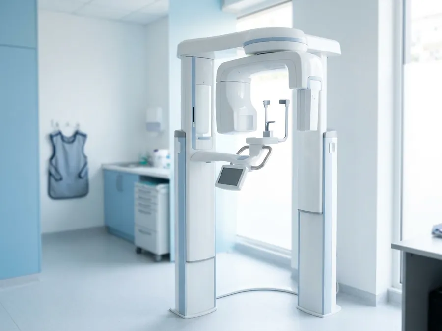
| Dental CT (CBCT) | Bone width and height, nerve and sinus position | Implant planning, impacted teeth, complex root canal cases |
Not every check-up needs an X-ray. How often you need one depends on your age, decay risk and symptoms, and is decided by the dentist.
How much radiation? Is it safe?
Dental X-rays are among the lowest-dose medical images. Roughly, a periapical film is a few microsieverts and a panoramic image is usually around 10–25 microsieverts. With dental CT, the dose depends on the size of the area scanned and usually ranges from tens to a few hundred microsieverts. For comparison, a person receives on average around 2,000–3,000 microsieverts a year from natural background radiation.
- Images aren't taken unless needed; bring any recent X-rays you have.
- CT is only requested when a two-dimensional image isn't enough.
- In children, the area and frequency are kept even more limited.
Can dental X-rays be taken during pregnancy?
Routine check-up X-rays can wait until after birth. But an X-ray needed to diagnose pain, an abscess or an emergency can be taken with appropriate protection; an untreated infection is also a risk for mother and baby. See dental treatment during pregnancy for more.
Why is a CT scan important before implants?
A panoramic image gives an idea of bone height but doesn't show its width. A CT scan shows bone width, the nerve canal in the lower jaw and the sinus in the upper jaw in three dimensions. This lets the implant's length and angle be planned more safely and shows in advance whether a bone graft or sinus lift is needed.
Imaging at Dent32 in Beylikdüzü
At Dent32 we show you the images taken or brought in on screen during the examination and explain the findings. We'll tell you at your appointment which image is needed and where it will be taken. Our dental clinic in Beylikdüzü is at Barış Mah., Ginza Lavinya, E-5 Yanyolu No:30 D:179, 34520 Beylikdüzü, Istanbul; to book, call +90 555 011 33 68.
Frequently asked questions
How often should dental X-rays be taken?
There's no fixed rule. Adults with low decay risk may only need bitewings every few years; with higher risk or symptoms, more often.
What's the difference between a dental CT and a panoramic X-ray?
A panoramic is a two-dimensional overview. A CT shows the bone in three dimensions and is used for implant planning and impacted teeth.
Can I bring an X-ray taken elsewhere?
Yes. If it's recent and clear, a new one may not be needed. A digital copy is easiest.